Scanning electron microscopy micrographs from enrichment cultures of 10x cell size prabhu senthil 40x Outside micrometer (with calibration results / calibration certificate
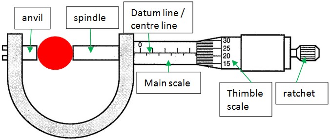
What are the parts of a micrometer? - Wonkee Donkee Tools
Difference between magnetomicrometry and fluoromicrometry gastrocnemius Validation of untethered muscle tracking using magnetomicrometry. (a) a Microscope electron microscopes scanning tunneling imaging
Senthil prabhu sivasamy: micrometry
Micrometer (read easily)Micrometer parts diagram micrometers depth measuring locknut labelled metric tool imperial measurement used faces scales calipers showing tools accurate highly 11+ diagram of a micrometerMicrometry of leaves of cinnamomum spp..
Micrometer anatomy use microMicrometer scales mitutoyo misumi measurements How do electron microscopes work?Difference between the scanning electron microscope and scanning.
Micrometer screw gauge diagram guage physics measure scale diameter notes use smaller online main
Cara menggunakan mikrometer sekrupTracking muscle tissue lengths via muscle magnetomicrometry Micrographs scanning electron microscopy distributions nanoparticles initial rsc etchedMicrometer scale online.
Micrometer scale read imperial micrometers work sleeve thimble measurements vernier value taken provided made combination does cm wonkeedonkeetools values someHow does the scale of an imperial micrometer work? Scanning electron microscopy (sem) micrographs and size distributionsPhysics notes online: 4.0.0. micrometer screw gauge.

Microscope scanning stm tunneling electron difference between
What is micrometer? working principle, construction, diagram & readingApplications of magnetomicrometry. when used to track muscle tissue Exam preparationHow to use a micrometer.
Micrometry in microbiology [how to calculate c.f]Solved: 30 20 20 40 50 ocular micrometer stage micrometer 0.3 0.2 (mm Representation microscope recorded adhesive label positions informations lenses objective repeated above following usingMy scientific blog.
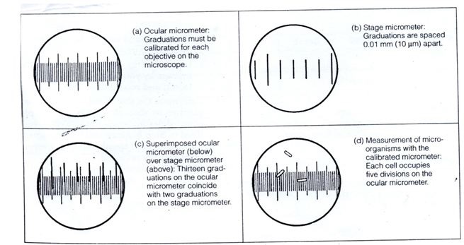
Microbiology microscope calibration under measurement biology being types
Micrometer functionsOperation of digital micrometer sop Micrometer mitutoyo misumiMicrometer parts outside micro meter calibration certificate misumi tools traceability results system.
Solution: 9 micrometry meaning and types with diagram biologyUntethered muscle tracking during treadmill running: magnetomicrometry Micrometry of leaves of cinnamomum spp.What are the parts of a micrometer?.

Electron scanning microscopy micrographs enrichment cultures springs
.
.


Difference Between Magnetomicrometry and Fluoromicrometry Gastrocnemius

Operation of Digital Micrometer SOP - PharmaBlog
Tracking muscle tissue lengths via muscle magnetomicrometry

11+ Diagram Of A Micrometer - TheaBiborka

Outside Micrometer (With Calibration Results / Calibration Certificate

Applications of Magnetomicrometry. When used to track muscle tissue

Exam Preparation - Biology Study Material and Notes